U WAVE IN ECG AND ITS ABNORMALITIES IN ECG | PPTX is a high-quality image in the Uci collection, available at 2048 × 1536 pixels resolution — ideal for both digital and print use.
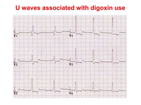
Learn the clinical significance of a U wave on an EKG. Discover how this subtle cardiac waveform relates to electrolyte imbalances like hypokalemia, bradycardia, or underlying heart disease. Our expert guide breaks down ECG interpretation, helping medical professionals and students identify U wave patterns accurately to improve diagnostic precision and patient cardiac monitoring outcomes.
Image Details
| Title | U WAVE IN ECG AND ITS ABNORMALITIES IN ECG | PPTX |
|---|---|
| Dimensions | 2048 × 1536 px |
| Category | Uci |
| Published | October 31, 2024 |
| Author | Zeus |
| Downloads | 1,105 |
| Views | 44 |
Frequently Asked Questions
This image has a resolution of 2048 × 1536 pixels. It is suitable for high-quality printing, digital presentations, and web use without losing clarity.
This image is part of the Uci collection. You can browse more images in this category to find similar content.
Click the Download button above the image to save it directly to your device. The image is provided in its original resolution of 2048 × 1536 px.
Yes! Scroll down to the More Images section below to explore related Uci images. You can also visit the full article for more context and a complete image gallery.
Read full article: U Wave Ekg