Understanding the Graphy Definition in Medical Context is essential for both practitioners and patients to ensure precise diagnosis and treatment. The concept of graphy, derived from the Greek word “grapho,” meaning “to write,” refers to various imaging and recording techniques utilized in modern medicine. Graphy processes enable healthcare professionals to visualize internal structures, facilitating accurate and effective medical interventions. This article delves into the definition and significance of graphy in a medical setting.
A graphy definition in medical terms involves the process of creating an image or record of an internal bodily function or structure. It relies heavily on technological advancements in imaging and recording to produce detailed visuals that can be used for diagnostic and therapeutic purposes. The term "graphy" encompasses a broad spectrum of techniques including, but not limited to, radiography, computed tomography (CT), magnetic resonance imaging (MRI), and ultrasound. Each method has distinct applications and advantages depending on the medical condition being examined.
Key Insights
- Primary insight with practical relevance: Graphy plays a critical role in early diagnosis, treatment planning, and monitoring of medical conditions.
- Technical consideration with clear application: MRI graphy, for example, provides superior soft tissue contrast, making it invaluable in neurology and orthopedics.
- Actionable recommendation: For practitioners, staying updated on the latest graphy techniques can enhance diagnostic precision and improve patient outcomes.
Radiography and Its Applications
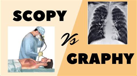
Radiography, commonly known as X-ray imaging, uses electromagnetic radiation to visualize the internal structures of the body. It remains a staple in various medical fields due to its efficiency and cost-effectiveness. Radiography is indispensable for detecting fractures, assessing bone density, and evaluating lung conditions such as pneumonia. The technique employs low doses of ionizing radiation, which pose minimal risk when appropriately managed.
MRI and Its Superior Contrast Resolution
Magnetic Resonance Imaging (MRI) employs a strong magnetic field and radio waves to generate detailed images of organs and tissues. Unlike radiography, MRI does not use ionizing radiation, making it particularly useful for examining soft tissues like the brain, spinal cord, and joints. The superior contrast resolution of MRI makes it a preferred method for diagnosing complex neurological disorders, including multiple sclerosis and certain types of tumors. Additionally, MRI provides comprehensive insight into cardiovascular structures, aiding in the diagnosis of heart and blood vessel conditions.
What are the primary risks associated with graphy techniques?
While graphy techniques are generally safe, ionizing radiation used in procedures like radiography and CT scans carries a slight risk of increasing cancer risk. However, this risk is significantly outweighed by the diagnostic benefits. To minimize risks, medical professionals adhere to the ALARA (As Low As Reasonably Achievable) principle to optimize radiation dose.
The evolution of graphy techniques continues to revolutionize the medical field, offering ever-improving tools for diagnosis and treatment. Mastery of these techniques not only enhances a healthcare provider’s diagnostic capabilities but also ensures better outcomes for patients. As the field of medical imaging progresses, staying informed about emerging technologies and best practices will be crucial for delivering top-notch medical care.